


Since childhood, we have become accustomed to running, jumping, boys like to climb and play football, girls are ropes and much more.And the active lifestyle enters the human mind so much that over the years when the muscle has been pulled somewhere, the joint has become ill somewhere, one does not even pay attention: "Well, think how many times Kolenko hurts."Here we speak in today's article, and why the knee can hurt and that it is always the usual result of sharp movement.
What is arthrosis?
Joint joint -A group of muscle system systems of different origins, but with similar biological, morphological and clinical manifestations.Their development is based on the degenerative lesion of all the components of the joint, primarily the cartilage, the thin bone, the synovial membrane, the ligaments, the capsules and the periarticular muscles, with the formation of marginal osteophytes and a clean or hidden, moderally pronounced synchronization.Because of this disease, pathological changes are captured by both cartilage and bone tissue.
Arthrosis is often called osteoarthosisand sometimes osteoarthritisOr
Statistics (epidemiology)
Of all diseases of the joints, arthrosis is up to 80% of cases.
The disease develops primarily in medium and old age.At a young age, arthritis, inflammatory processes and muscle bone system can occur with congenital pathology.
The symptoms of arthrosis X rays are detected in most people over 65 and nearly 95%.
Women suffer almost twice more in arthrosis than men.The occurrence rate increases during postmenopause.
Hereditary factors play a major role in the development of arthrosis.It has been found that the frequency of developing the disease in the family of patients with osteoarthritis is twice higher than the population, and the development of arthrosis is increased 7-8 times.
Arthrosis - ICD
- MKB-10: M15-M19, M47
- MKB-9: 715
- MKB-9-KM: 715.3
The joint symptoms (clinical picture)
The clinical manifestation of the disease and their severity depend on the localization of the pathological process, the health of the patient and the image of life.
The first signs of joint
Arthrosis often begins gradually, imperceptibly for the patient.
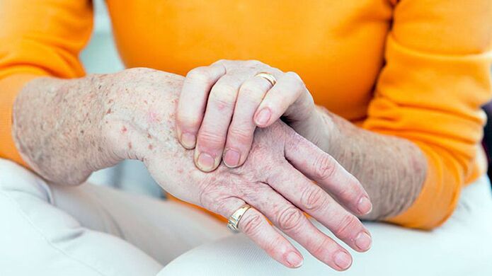
The first symptom of the disease is usually a short, minor joint pain (arthralgia) that bears the highest load.These are primarily the joints of the lower limbs-the knees, the hips, the plus-phalanx joints with the front thumb of the foot.The upper limb, interfalngeal joints, the brush thumb, are more commonly affected by the carpal pattern.
Arthrosis usually begins with one joint injury, but after a while other joints are involved in the process.
The joint symptoms
In the case of arthrosis, patients complain of pain, crisis, restriction of joint movement, swelling and joint deformation.
Separate -it is worth living in the nature of the pain.Mechanical and initial pain is possible in arthrosis.Mechanical pain occurs with the load on the affected joint.Such pain is primarily a disorder at rest and disappears after several hours of rest.The appearance of this type of pain results in a gradual increase in bone pressure during physical effort.The pressure causes bone rays and irritation of painful bone tissue.
Starting pain appears at the beginning of the walk, then stops quickly and occurs again during physical effort.Starting pain can occur by friction of the joint surfaces of the affected joint.Small particles of necrotic cartilage fall on the cartilage.In the first steps, these particles are pushed into the cavity of the joint bag and the pain disappears.
In the case of arthrosis, the pain can be associated with parallel inflammation and momentum (inflammation of the soft periarticular tissues, leagues and joint bags).This pain only occurs in movements in which the affected tendons are involved and in certain situations of the joint while moving.
Pathological changes usually begin with large joints, which are subjected to great physical effort throughout the day.At the beginning of the disease, pain is the result of inconsistency of the microcirculation channel and the needs of the joint tissues.Therefore, patients slowly take the first few steps to reduce pain and only accelerate the pace of walking.The pain can occur after half -hour walk or work in a constant position.This is a signal to change the type of load, short -term relaxation or work type.
At later stages of the disease, arthralgia may be minimal load on the joint and may remain at rest for a long time.The reason for this is that in later stages, rough changes in the joint tissues, the destruction of the articular cartilage and the secondary synovitis develop.In the bone -cheese tissues, with the formation of huge, severe changes, individual fragments can be separated and may fall into the joint and cause sharp pain.This phenomenon is called the symptoms of the joint mouse.
Deformation is noteworthy when examining joints.In addition, in the case of arthrosis, periastal soft tissue, regional muscles hypotrophy, and the displacement of the limb axis thicken.The thickening of the interalAngeal joints with bone growth and sealing of periarticular tissues are called Gerberden nodes.
When the joint feels, the pain is localized in the arthritis, in the recording points of the joint capsule, but this symptom of the disease is not always.The swelling and pain of the joint is determined by secondary synovitis.
Violation of joint function is manifested by limiting the amplitude of movements during the early stages of arthrosis.This is due to injury to periosematic tissues and synovitis.
In later stages of the disease, clinical manifestations of contractures are different in terms of severity.Most often, the functions of the knee and hip joints are damaged.
Symptoms of arthrosis depending on the localization of pathology
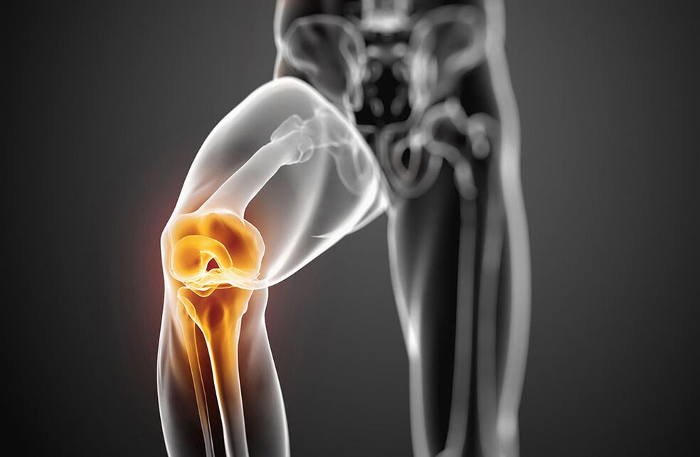
Arthrosis with damage to knee joints - symptoms
The lesion of the knee joints with arthrosis is called gonarthrosis.Primary gonarthrosis develops in menopause in women.Secondary causes are most commonly injured in the knee joint and violation of static with curvature of the spine and flat legs.Patients complain of the pain of the knee joint, which occurs during movements, especially when walking up the stairs.The pain is localized to the front or inside of the knee joint.The joint movement is limited: first bending and later extension.A crunch often occurs when mobility.With the development of reactive synovitis, the pain increases and is concerned at rest.We determine the swelling of the joint, the pain during palpation, the redness (hyperemia) and the increase in skin temperature.Over time, bone growth due to the growth of the knee joints occurs.
Arthrosis with the damage to the hip joints - symptoms
The lesion of the hip joints is called coksartrosis.This is the most serious form of arthrosis.The causes of the disease may be the congenital dysplasia of the hip joints, injuries, and menopause.Patients have pain in the joints during movements in a constant position.The restriction of the joint movement is gradually increasing (first internal and external rotation, later bending).There is lame to shorten the limb.In the case of bilateral damage, duck walking is typical.The atrophy of the thighs and the muscles of the buttocks develops.The joints do not swell with cartrosis.Determine the limited pain of the femur head.
At the initial stage of arthrosis, joint functions remain.With the further development of the disease, it is first temporarily limited, and then the ability to work is completely lost, the patient loses the ability to self -care and needs external help.
The joint reasons
Arthrosis is based on the primary degeneration of the articular cartilage, accompanied by the devastating changes of the joints that make up the joints.Such degeneration occurs as a result of mechanical loads on the joint surface of the cartilage and the chances of load compensation.
Many factors can participate at the same time when developing degenerative changes in joint cartilage:
- Functional overloads, including professional, household and sports, cartilage mycotrauma;
- joint injuries;
- Infectious and non -specific inflammation of the joint;
- common dysplasia leading to a violation of comparison of joint surfaces;
- Violation of body statics as a result of curvature of the spine (kyphosis, skoliosis, pathological lordosis, etc.), flat legs;
- Chronic hemarthrosis:
- Diseases with metabolic disorders (gout, obesity, chondrocalcinosis);
- Osteodistrophy or Pedget;
- osteomyelitis;
- The pathology of the peripheral nervous system with the loss of sensitivity;
- Endocrine pathology (acromegalia, diabetes, amenorrhea, hyperthyroidism);
- Hereditary trend.
The risk factors of arthrosis include elderly, women's sex, and obesity.
Development mechanism
The metabolism in the cartilage is based on quantitative and qualitative changes in the main substance of the cartilage.The main substance consists of proteoglycans that provide collagen stability.The development of arthrosis is accompanied by insufficient formation or increased destruction of cartilage components.
In the cartilage tissue, in the case of osteoarthritis, the content of hyaluronic acid, chondroitin and keratin is reduced.In addition, changed proteoglycans lose water retention.It is absorbed by a collagen that swells and caused by a reduction in cartilage resistance.
If the chondrocites are damaged, they begin to produce collagen and proteoglycans, which are not typical of normal cartilage tissue.These altered materials cause the biochemical properties of the cartilage.
Immune deficiency is very important in the development of arthrosis.The destruction of cartilage proteoglycans is accompanied by the appearance of cellular and humorous immune reactions.This, in turn, causes progressive fibrosis and sclerosis to the pathological changes of the synovial membrane, intraarticular synovial fluid and cartilage violation.A lower -level synovial shell supports the progress of the degenerative changes in the articular cartilage.
The hereditary factor has a certain value in the formation of arthrosis.
The joint classification
Arthrosis is divided into two groups: primary and secondary.
Distribution (primary arthrosis):
- Local (damage to three joints)
- Generalized or generalized polyarthrosis (defeat of three or more taste).
Depending on the destination (secondary):
- A. Tasobed joint (cokesartrosis);
- A. the knee joint (gonarthrosis);
- A. The elbow joint;
- A. the shoulder joint;
- A. spine;
- A. Cervical class (unkoarthrosis);
- A. hands;
- A. ankle joint (crusartrosis)
- A. Stop.
Etiology:
- post -traumatic
- metabolic
- Because of endocrine pathology.
The joint diagnosis
The diversity of clinical manifestations and variants of arthrosis makes it difficult to diagnose the disease.The antiquity of the diagnosis also results in a lack of specific symptoms and the hidden appearance of the disease.It is very important to determine the factors that contribute to the development of arthrosis:
- chronic joint trauma;
- Long -term execution of stereotypes;
- physical activity on the joint for a certain period of time;
- violation of the metabolism of salt or fat;
- Hereditary wickedness of the muscle bone system.
The X -Gay test is the most important meaning in diagnosis of arthrosis.The visual radiography of both knee joints is performed in a direct position, in a bent position, also sideways.The classic signs of X -rays are as follows: narrowing of joint gap, presence of osteophytes, subchondral bone sclerosis and subchondral cysts.There are the following stages of radiological changes in arthrosis:
- 0 - No change.
- I - Dubious signs from a radiological point of view.
- II. - minimal changes (slight narrowing of the joint gap, subsidiary osteosclerosis, single osteophytes).
- III. - Moderate manifestations (moderate reduction of charts, multiple osteophytes).
- Arc.- Expressed changes (the joint gap is not visible, more rough osteophytes are determined), synovitis is often present.
In the presence of these symptoms, no additional tools are required.
Their lack or low severity, joints, MRI, scintigraphy occur.
Clinical examination of blood, urine and intraarticular synovial fluid is not included in the list of mandatory tests to diagnose arthrosis.But these tests are needed to exclude such joint pathologies.
The joint clinical and diagnostic symptoms:
- Mechanical joint pain;
- fatigue;
- feeling of instability on the joints of the lower limbs;
- damage to the joints of the leg and the front fingers of the hands;
- gradual appearance of the disease;
- slow progressive current;
- joint deformation;
- Regional muscles hypotropy;
- Returning synovitis;
- Restriction of movement in the joint;
- X -Ray changes.
Arthrosis should be distinguished by damage to rheumatoid arthritis, infectious, metabolic and reactive arthritis.
Unlike arthrosis, rheumatoid arthritis begins with inflammation of the small joints of the hands and feet.This is characterized by the inflammatory type of intense pain, the morning stiffness of the joints, and the presence of rheumatoid lumps.
Gotric arthritis is primarily found in men.The first plus-phalanx joint of the leg thumb is characterized by high local activity with acute paroxysmal pain.In addition to gout, the presence of the tofus is characterized by the X -ray.
Psoriatic joint inflammation of the skin, especially the scalp, spindle deformation of the fingers and the bright raspberry color over the affected joints.
Infectious joint inflammation is characterized by an acute beginning, rapid development and path, sharp pain, high temperature and effectiveness of antibacterial treatment.
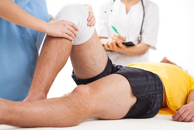
Treatment of joint treatment
Joint treatment should be long, complex.Principles of treatment of arthrosis:
- Unloading of the joints (the right way to mobility and mechanical loads, dosage, reduction of body weight, exclusion of long-term positions, weights, strengthening the muscle-ligament device using physiotherapeutic exercises, massage, electrical stimulation).
- Conservative correction of static disorders (the use of orthopedics, corset, supervisors).
- The effect of general metabolism and blood circulation (the use of biostimulants, vasodilizing drugs, balneotherapy and physiotherapy courses twice a year).
- Elimination of reactive synovitis, anti -inflammatory treatment.
Patients in the arthritis show a diet, which is limited by limiting salt, sugar, strong tea, coffee, smoked meat, and sharp foods.This improves the sensitivity of vascular and joint receptors, restores the sound of blood vessels, and normalizes the replacement in the chondrocytes.In the case of arthrosis, drink sufficient fluid (at least 8 glasses of water daily).
Medication for arthrosis includes the use of rapid, anti -inflammatory and analgesics (non -sertoidal anti -inflammatory drugs -NSAIDs), basic drugs -chondroprotectors.Not-?Non-selective and selective TSO-2 inhibitors are used from NSAIDs.
NSAIDs are used as an ointment or gel as the local therapy of the affected joints.
In the presence of reactive synovitis, tendinitis or endovaginitis, when treatment of NSAIDs is ineffective, adequate intraarticular or intramuscular corticosteroids.
Condroprotectors (chondroitin, glucosamine, hyaluronic acid) are used to prevent degeneration of articular cartilage.
Treatment of chondroprotectors is indicated by I-III in joint clinical and radiological stages.
In addition to direct chondroprotectors, drugs that stimulate the restoration of cartilage tissue (biogenic stimulants).These drugs are used during remission, in the absence of reactive synovitis.
In the case of arthrosis, drugs that improve microcirculation are also indicated.Varicosis of the lower limbs requires a correction of venous blood flow.
Patients with arthrosis require timely diagnosis and treatment of osteoporosis.
Physiotherapy of arthrosis
Treatment physical methods are also related to the basic therapy of arthrosis.During their influence, microcirculation of metabolic processes, blood and tissue fluid is stimulated and is restored by neurogumoral regulation.
The complex of the treatment of arthrosis includes inductoria, microwave therapy, pulse currents, electrophoresis of drugs and magnetic therapy.To eliminate synovitis, ultraviolet irradiation of the affected joints is used in erythhemum doses, the ultra -high -frequency electrical field, the electrophoresis of analgin, dimexide or hydrocortisone.
To prevent the progression of arthrosis, it is recommended to reduce body weight, increased joint loads of joints, walking in the oppressed area, increased humidity and hypothermia.The unique selection of shoes and supervisors is important.
Gonarthrosis, regular physical exercises, swimming and cycling strengthen the muscles.Department of heavy and light athletics, football is not recommended.
Therapeutic exercises are performed differently, in a sedentary position, lying in the pool.Movements should not be intense, traumatic, and the number and number of repetitions are gradually increasing, avoiding overloads.
Popular and effective methods of treating arthrosis include massage and kinin -natherapy.
With significant changes in the joints by deformation, mobility is recommended and surgical treatment.Artroplasty, endoprostics, osteotomy are performed.
The prognosis of the disease
Primary arthrosis rarely leads to total disability.In the presence of reactive synovitis, patients become temporarily disabled and sometimes have to change the profession.In the case of secondary cocarchy, prognosis is less favorable due to the rapid progressive process of the disease, which results in significant damage to joint functions.In such cases, disability can occur during several years of illness.
Preventing a joint
Primary joint prevention should begin in childhood.This is the following:
- Prevention and treatment of skoliosis;
- Correction of flat legs with special supervisors;
- physical education classes to strengthen muscles and ligaments;
- Prevention of reasonable nutrition and metabolic disorders;
- Limit heavy sports in childhood and adolescence;
- alternating work at a sitting table walk;
- Organizing the workforce and other employees for businesses with heavy physical activity.
Secondary prevention provides measures that prevent recurrent reactive synovitis.These include dosage walking, restriction of physical effort, support and fitting other measures.In the case of severe symptoms of arthrosis, basic medicines should be continuously taken.General strengthening therapy, blood circulation improvement and metabolism, and annual treatment are recommended.
Which doctor will you go to?
- Rheumatologist
- Orthopedic